Automatic mitosis detection in breast cancer tissue sections
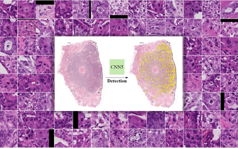
Manual counting of mitotic tumor cells in tissue sections constitutes one of the strongest prognostic markers for breast cancer. This procedure, however, is time-consuming and error-prone. David Tellez et al developed a method to automatically detect mitotic figures in H&E stained breast cancer tissue sections based on convolutional neural networks (CNNs). The system was trained in a single-center cohort and evaluated in an independent multicenter cohort from The Cancer Genome Atlas on the three tasks of the Tumor Proliferation Assessment Challenge (TUPAC). The image shows a selection of patches identified by the CNN as containing a mitotic figure. From 181 detections, 128 patches were classified as true positives by a resident pathologist, resulting in a precision score of 0.707. The work was published in Transactions on Medical Imaging.
← Back to overview